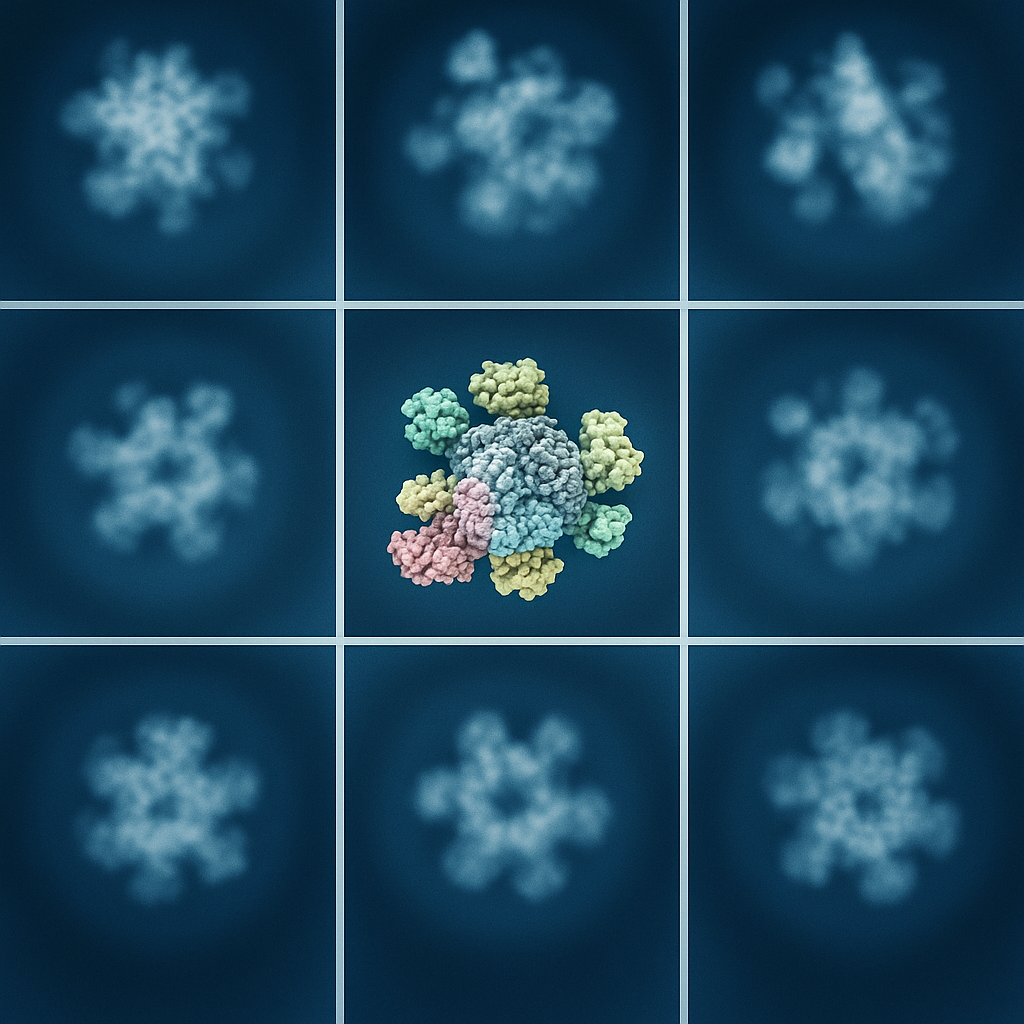
Bridging Cryo-EM & Molecular Dynamics
How does a structure become a mechanism?
Cryo-electron microscopy reveals the architecture of biomolecular assemblies, often in several distinct conformations. The transitions between those structures and the interactions that stabilize them are central to biological function. We use molecular dynamics and coarse-grained models to connect structural observations to motion, energetics and molecular mechanism.
In this collaborative approach, experimental structures constrain the models, simulations identify interactions and accessible transitions, and experimental measurements test the resulting explanations. Our contribution centers on computational modeling and interpretation of molecular dynamics. Cryo-EM, NMR and single-molecule measurements are supplied by the relevant experimental collaborators.
Related studies: Costa, Gur, Krieger and Bahar (2024), Computational biophysics meets cryo-EM revolution in the search for the functional dynamics of biomolecular systems; Ferro et al. (2022), Structural and functional insight into regulation of kinesin-1 by microtubule-associated protein MAP7; Hansen et al. (2025), Structural basis of Pseudomonas biofilm-forming functional amyloid FapC formation.
Microtubule regulators: architecture explains selectivity
Our MAP7 study illustrates how structural and dynamic information can explain apparently competing effects of a regulatory protein. Cryo-EM defined its microtubule-bound organization, while simulations and functional measurements helped connect that organization to kinesin-1 recruitment and movement. This work makes the relationship between a binding footprint and a transport phenotype explicit.
The MAP9 preprint extends this reasoning to a different architecture. Its helical binding mode around the microtubule helps explain how a regulator can stabilize the lattice while allowing some motors to pass and obstructing others. The latest public version is a preprint, so its findings are identified separately from the peer-reviewed MAP7 study.
Related studies: Ferro et al. (2022), Structural and functional insight into regulation of kinesin-1 by microtubule-associated protein MAP7; Cetin et al., Structure and mechanism of microtubule stabilization and motor regulation by MAP9. bioRxiv preprint, version 2, August 25, 2026.
FapC: from a disordered protein to a biofilm scaffold
Our 2025 FapC collaboration combined solution NMR, a roughly 3.3-angstrom cryo-EM fibril structure and all-atom simulations. FapC is a functional amyloid protein that contributes to the biofilm matrix of Pseudomonas. The experimental data describe different stages of its structural organization, while simulations connect changes in local secondary structure to the larger rearrangements required for assembly.
The simulations compared isolated subunits with subunits incorporated into fibrillar assemblies. They showed why the molecular environment matters: a conformation observed in a fibril is stabilized by interactions that an isolated monomer does not possess. The combined study provides a mechanistic account of assembly and disassembly, and a structural foundation for investigating biofilm-associated infection.
Related studies: Hansen et al. (2025), Structural basis of Pseudomonas biofilm-forming functional amyloid FapC formation.

Connecting states without inventing the path between them
A model fitted to a density map and a trajectory generated from that model answer different questions. We distinguish experimentally resolved features from simulation-derived motions and test whether proposed transitions are consistent with the available evidence. Enhanced sampling can explore transitions beyond those observed in a conventional trajectory, but the resulting pathway must be interpreted in light of the method and its assumptions.
This distinction is essential when studying partly disordered proteins, transient interfaces and large assemblies. Our goal is a molecular explanation that joins complementary observations while making clear which features are measured and which are predicted.
Related studies: Costa, Gur, Krieger and Bahar (2024), Computational biophysics meets cryo-EM revolution in the search for the functional dynamics of biomolecular systems; Gur, Madura and Bahar (2013), Global transitions of proteins explored by a multiscale hybrid methodology: Application to adenylate kinase; Das et al. (2014), Exploring the conformational transitions of biomolecular systems using a simple two-state anisotropic network model.
